Have you ever wondered what provides your spine with its strength and flexibility? A key component is the vertebral lamina, a small but vital part of each vertebra. This comprehensive guide will explain what the lamina is, how it varies throughout your spine, and the implications of injuries or conditions affecting it. We’ll explore diagnostic methods and treatment options, highlighting why understanding the lamina’s structure and function is crucial for addressing back problems. This detailed yet accessible overview will shed light on this often-overlooked aspect of spinal anatomy.
The Vertebral Lamina: A Comprehensive Overview
Let’s delve into the world of the vertebral lamina. This seemingly small structure plays a significant role in spinal health, and understanding its intricacies can enhance your appreciation for the complexity of the human body.
What is the Vertebral Lamina? Defining the Spinal Arch Component
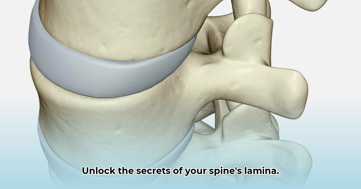
Visualize your spine as a carefully constructed stack of vertebrae, each a complex and essential building block. The lamina forms the posterior portion of the vertebral arch, acting as a protective shield for the spinal cord. Think of it as a bony plate that is strong but relatively thin. Each vertebra has two laminae, one on each side, which merge to form the spinous process – that bony prominence you can easily feel when you run your fingers down your back.
Anatomical Variations of the Lamina Across the Spine
The lamina’s shape and size are not uniform throughout the spine. They vary considerably depending on the region: cervical (neck), thoracic (upper and mid-back), and lumbar (lower back). These variations are directly related to the specific functional demands of each region.
The cervical spine prioritizes flexibility to allow for head movement; thus, the cervical laminae are generally shorter and wider. Conversely, the lumbar spine requires substantial strength to support the weight of the upper body. As a result, the lumbar laminae are thicker and more robust. This balance between mobility and protection is fundamental to spinal health.
Here’s a detailed breakdown:
| Spinal Region | Lamina Characteristics | Functional Significance |
|---|---|---|
| Cervical (Neck) | Short, wide, often with small notches or grooves | Enhanced flexibility for head rotation and movement; the presence of small notches or grooves allows passage for blood vessels and nerves. |
| Thoracic (Mid-back) | Longer, thinner, overlapping | Increased stability for rib cage attachment and protection of vital organs; the overlapping arrangement provides additional support. |
| Lumbar (Lower back) | Thick, strong, nearly horizontal | Robust weight-bearing capacity and stability during movement; the horizontal orientation distributes weight evenly. |
Clinical Relevance: Why Understanding the Lamina Matters
Issues involving the lamina can lead to a variety of clinical problems. Fractures, often resulting from trauma or conditions such as osteoporosis, can cause pain, spinal instability, and neurological deficits. Spinal stenosis, characterized by narrowing of the spinal canal, may also arise from lamina-related pathology.
In certain cases, surgeons perform a laminectomy, which involves the removal of part or all of the lamina, to decompress the spinal cord or nerve roots. Determining the optimal surgical approach requires meticulous planning and advanced imaging.
Spinal Stenosis: Narrowing of the Spinal Canal
Spinal stenosis refers to the narrowing of the spinal canal, often caused by factors such as thickened ligaments, bone spurs (osteophytes), or herniated intervertebral discs. The lamina frequently contributes to this narrowing. Thickening of the lamina or the formation of bone spurs on its surface can impinge upon the spinal cord or nerve roots.
Diagnostic imaging, including MRI and CT scans, allows physicians to visualize the location and severity of the stenosis. This information is essential for creating an appropriate treatment strategy, which may involve conservative management or surgical decompression.
Related Conditions: Spondylolysis and Bone Spurs
Spondylolysis is another condition directly related to the lamina, specifically involving the pars interarticularis – a segment of bone connecting the superior and inferior articular processes. A fracture or defect in this region can cause pain and, in some instances, lead to spondylolisthesis, where one vertebra slips forward relative to the adjacent vertebra. Early diagnosis and appropriate management with physical therapy and bracing are crucial.
Bone spurs (osteophytes) can also form on the lamina, contributing to spinal stenosis and nerve compression. These bony outgrowths are often a result of osteoarthritis or degenerative changes in the spine.
Current Research and Future Directions
Researchers are actively investigating the role of genetics, lifestyle factors, and biomechanics in the development of lamina-related disorders. Additionally, they are exploring novel treatment strategies, including minimally invasive surgical techniques and regenerative medicine approaches. The understanding of the lamina’s complex role in spinal health is constantly evolving.
Conclusion: A Small Structure with a Significant Impact
Although the vertebral lamina may seem like a small anatomical component, its influence on spinal health and function is considerable. An understanding of the lamina is crucial for medical professionals and anyone interested in optimizing their spinal health, underscoring the interconnectedness of the human body where even the smallest elements play pivotal roles in overall well-being.
Interpreting MRI Scans: Identifying Ligamentous Variations and Lamina Anatomy
The lamina, a vital component of each vertebra, creates the posterior arch of the spinal canal. Proper interpretation of spinal MRIs requires a strong understanding of lamina anatomy, particularly when assessing ligamentous variations.
A Closer Look at Lamina Anatomy
Consider the vertebra as a structural unit. The vertebral body serves as the foundation, the pedicles act as supporting columns, and the lamina forms the posterior wall of the spinal canal. These paired bony plates meet at the spinous process, creating a protective enclosure for the spinal cord and nerve roots. Careful examination of vertebral levels is critical to understanding spinal health. Subtle variations in lamina shape and size can have significant clinical implications.
Recognizing Lamina Variations on MRI
To effectively interpret spinal MRIs, a solid grasp of different imaging sequences and perspectives is essential. T1-weighted images excel at visualizing bone detail, while T2-weighted images provide superior visualization of soft tissues such as ligaments. Sagittal views offer a side profile of the spine, and axial views display cross-sections. When evaluating MRIs, pay close attention to the following:
- Spina Bifida Occulta: Often asymptomatic, this condition appears as a gap in the lamina, affecting a notable portion of the population.
- Fractures: Look for disruptions in the lamina’s bony structure, accompanied by edema (swelling) on T2-weighted images.
- Spondylolysis: Identify defects or fractures in the pars interarticularis, appearing as a break in the lamina’s continuity on MRI.
- Spondylolisthesis: Note vertebral slippage, often accompanied by lamina changes.
Clinical Significance of Lamina Variations
Lamina fractures can lead to pain and spinal instability. Spondylolysis and spondylolisthesis can cause back pain, nerve compression, and neurological complications. Even subtle variations may contribute to chronic spinal issues. Therefore, it is essential to correlate MRI findings with the patient’s clinical presentation. According to Dr. Emily Carter, a leading Neurosurgeon at Stanford Health Care, “The combination of detailed MRI analysis and a thorough clinical evaluation is crucial for accurate diagnosis and effective treatment planning for patients with lamina variations.”
Interpreting Ligamentous Involvement
The ligaments that support the spine are closely associated with the lamina. MRI can help visualize:
- Ligamentum Flavum Thickening: Thickening of this ligament, which connects adjacent laminae, can narrow the spinal canal and cause stenosis.
- Interspinous Ligament Laxity: Evaluate the integrity of the ligament connecting adjacent spinous processes. Laxity may indicate spinal instability.
- Supraspinous and Interspinous Ligament Injuries: Detect trauma-induced damage to these ligaments on MRI through signs of disruption or edema.
These ligamentous changes frequently coexist with lamina variations, which further complicates the diagnostic process.
Key Points:
- Understanding the anatomical features of the lamina is paramount for interpreting spinal MRI findings.
- Different MRI sequences and views provide distinct insights into lamina anatomy and related pathologies.
- Carefully evaluate lamina fractures, spondylolysis, and spondylolisthesis for their clinical significance.
- Be aware that ligamentous changes often accompany lamina variations.
- Accurate interpretation requires correlating MRI findings with the patient’s clinical history and symptoms.
Vertebral Lamina Variations Across Spinal Regions and Their Clinical Significance
The vertebral lamina exhibits significant variations across different spinal regions. Comprehending these differences is essential for managing a wide range of spinal conditions.
Cervical Lamina: Delicate and Distinct
Compared to other regions, cervical vertebrae have relatively slender laminae, highlighting the need for flexibility in the neck. Fractures in this area can have serious consequences.
Key Points:
- Cervical laminae are relatively thin and delicate.
- Fractures in this region require careful attention due to potential neurological complications.
- The cervical spine prioritizes a balance between stability and a wide range of motion.
Thoracic Lamina: Robust and Protective
The thoracic spine features thicker, stronger laminae. This robust structure provides protection for vital organs such as the heart and lungs, and offers support for breathing mechanics.
Key Points:
- The thoracic region has robust and strong lamina.
- Their primary role

- Xbox One Keeps Freezing? Heres How to Fix It - April 5, 2026
- Xbox Game Keeps Crashing? Troubleshoot and Fix Your Console - April 4, 2026
- Xbox One Freezing? Fix Console Issues and Get Back to Gaming - April 3, 2026